A-Eye Web Platform
Automated 3D MRI Segmentation of Human Eye Structures
Upload a human T1-weighted orbital MRI and receive fully-automated segmentation masks for
9 structures of the eye, together with quantitative biomarkers —
axial length and volumetry — delivered as a ready-to-use .zip archive.
What does A-Eye segment?
The pipeline is based on nnU-Net and was trained on a large-scale, multi-centre cohort of healthy adult subjects. It provides segmentation masks and quantitative biomarkers for the following structures:
 Lens
The crystalline lens
Lens
The crystalline lens
 Globe
The full eyeball volume
Globe
The full eyeball volume
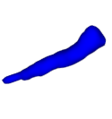 Optic Nerve
The optic nerve trunk
Optic Nerve
The optic nerve trunk
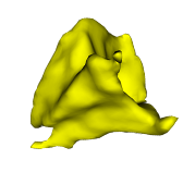 Intraconal Fat
Fat inside the muscle cone
Intraconal Fat
Fat inside the muscle cone
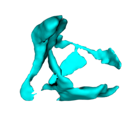 Extraconal Fat
Fat outside the muscle cone
Extraconal Fat
Fat outside the muscle cone
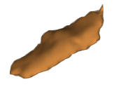 Lateral Rectus
Lateral rectus muscle
Lateral Rectus
Lateral rectus muscle
 Medial Rectus
Medial rectus muscle
Medial Rectus
Medial rectus muscle
 Inferior Rectus
Inferior rectus muscle
Inferior Rectus
Inferior rectus muscle
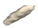 Superior Rectus
Superior rectus muscle
Superior Rectus
Superior rectus muscle
Pipeline Overview
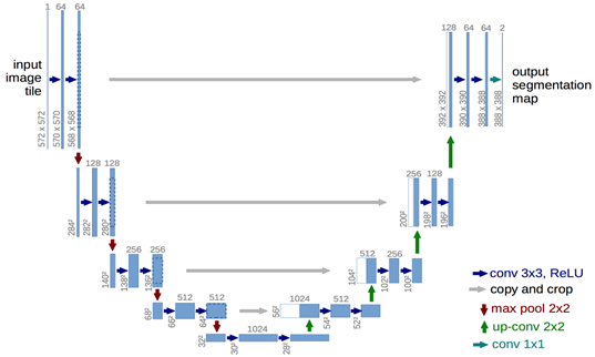
A-Eye uses nnU-Net, a self-configuring deep-learning framework for biomedical image segmentation. The model was validated on 1,245 healthy adult subjects and achieves state-of-the-art performance across all nine orbital structures.
Accepted input formats
.nii— NIfTI image.nii.gz— compressed NIfTI.zip/.7z— archive of NIfTI or DICOM files.dcm— DICOM (inside an archive)
T1-weighted sequences only. Up to 10 cases per submission.
What you get back
- Per-eye masks — left, right, merged (
.nii.gz) - Cropped per-eye masks used for the biomarkers
- Inference details and the logs of your run
- Axial length visualisations (
.png) - Biomarkers summary table (
biomarkers.csv)
All results packaged in a single .zip download, together with the license agreement. Each case folder also holds the image the masks are defined on, so you can check the overlay straight away — keep your own copy of what you uploaded.
Cite this work
If A-Eye contributes to your research, please cite the associated publication and dataset:
Jaime Barranco, Adrian Luyken, Hamza Kebiri, Philipp Stachs, Pedro M. Gordaliza, Oscar Esteban, Yasser Aleman, Raphael Sznitman, Oliver Stachs, Sönke Langner, Benedetta Franceschiello, Meritxell Bach Cuadra. A-eye: Automated 3D MRI Segmentation and Morphometric Feature Extraction for Eye and Orbit Atlas Construction. PLOS ONE. https://doi.org/10.1371/journal.pone.0352257
Barranco J, Luyken A, Stachs P, Esteban O, Aleman-Gomez Y, Stachs O, et al. MR-Eye atlas: a large-scale atlas of the eye based on T1-weighted MR imaging [dataset]. Zenodo; 2024. https://doi.org/10.5281/zenodo.13325369
Resources & downloads
All code, eye atlases, and pretrained model weights behind A-eye are openly available.